

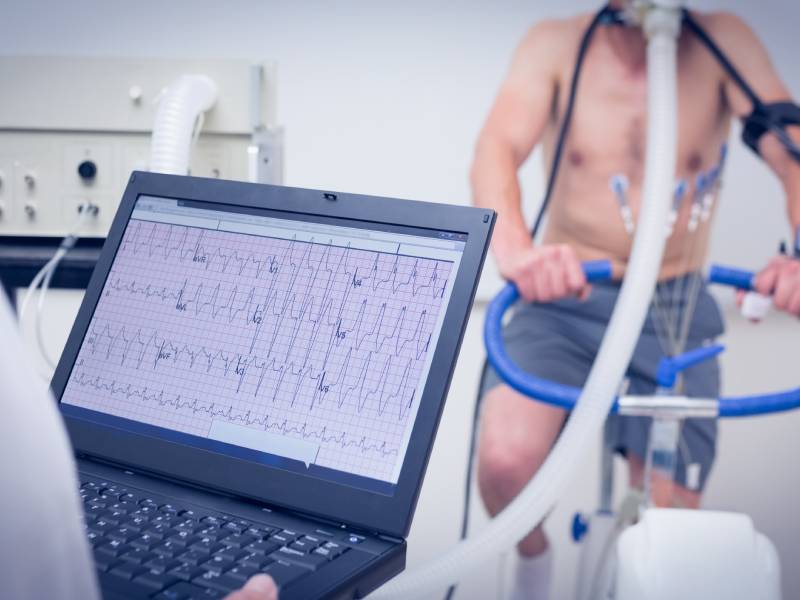
An ECG and an echocardiogram are two of the heart tests doctors use most often, and their names are easy to mix up. Both are painless and neither uses X-rays, but they answer different questions. An ECG (electrocardiogram) records the heart’s electrical activity: how fast and how regularly it beats. An echocardiogram is an ultrasound scan of the heart: it shows how the heart is built, how well it pumps and how the valves work.
At Aura Medical Clinic in Tirana, Prof. Dr. Albi Fagu, Head of the Vascular Surgery Department at “Shefqet Ndroqi” Hospital, offers cardiology consultations. His background is in cardiac surgery, with 2 years of experience in Belgium and 10 years at Universitätsklinikum Freiburg. ECG and echocardiography are both part of the clinic’s cardiology service. This article explains what each test shows, how it is done and when a doctor suggests one, the other or both.
ECG or echocardiogram: a comparison
| What to compare | ECG (electrocardiogram) | Echocardiogram (heart ultrasound) |
|---|---|---|
| What it records | The electrical signals that make the heart beat. | Moving pictures of the heart, made with ultrasound. |
| Main question it answers | How is the heart beating: rate and rhythm? | How is the heart built, and how well does it pump? |
| How it is done | About 10 small sticky electrodes are placed on the chest, arms and legs while you lie still. | Gel is put on the chest and a small probe is moved over the skin while you lie on your left side. |
| How long it takes | A few minutes; the recording itself takes seconds. | Usually between 15 minutes and an hour. |
| Does it hurt? | No. Removing the electrodes can pull on body hair. | No. The gel feels cold and the probe presses lightly on the chest. |
| Preparation | Usually none; do not put body lotion on your chest that day. | Usually none for the standard test. |
| What it does not show | Rhythm problems that come and go may not appear during the short recording. | It does not record the rhythm over time and does not show the coronary arteries (the heart’s own arteries) directly. |
The two tests complement each other: one does not replace the other. Which one you need depends on your symptoms, the examination and your medical history, and the doctor decides this with you.
What an ECG shows
Every heartbeat starts with an electrical signal. An ECG records this signal through the electrodes on the skin and draws it as a series of waves. From these waves the doctor can see:
- whether the heart beats too fast, too slowly or irregularly, for example in atrial fibrillation;
- whether the electrical signal is delayed on its way through the heart;
- signs that part of the heart muscle is not getting enough blood, or was damaged by an earlier heart attack;
- signs that may point to a thickened heart muscle, which an echocardiogram can then measure.
A standard ECG captures only a short moment. If palpitations or dizziness come and go, the ECG can be normal between episodes. In that case the doctor may suggest a longer recording, for example over 24 hours or more, with a small portable device.
What an echocardiogram shows
An echocardiogram uses the same ultrasound technique as other ultrasound scans to make moving pictures of the heart. It shows:
- the size of the heart chambers and the thickness of the heart walls;
- how strongly the heart muscle contracts, and whether some areas move less, for example after a heart attack;
- how the four heart valves open and close, and whether a valve is narrowed or leaks;
- the direction and speed of blood flow inside the heart (Doppler);
- fluid around the heart.
The report often gives the ejection fraction (EF): the percentage of blood that the main pumping chamber of the heart pushes out with each beat. Ask the doctor what your value means for you.
When a doctor suggests which test
An ECG is often the first test, because it is fast and simple. It is commonly done for:
- palpitations, or a racing or irregular heartbeat;
- chest pain, shortness of breath, dizziness or fainting;
- high blood pressure, diabetes or other risk factors for heart disease;
- a check before an operation or before competitive sport, where this is required;
- follow-up of a known heart condition, or of medicines that can affect the heart rhythm.
An echocardiogram is usually added when the doctor wants to look at the structure and pumping of the heart, for example:
- when a heart murmur is heard with the stethoscope;
- with shortness of breath or swollen ankles that may come from the heart;
- when heart failure or valve disease is suspected, or to follow them up;
- after a heart attack;
- when the ECG shows changes that need a closer look, such as signs of a thickened heart muscle;
- when close relatives have had a disease of the heart muscle (cardiomyopathy) or a sudden cardiac death at a young age.
Can a normal ECG rule out heart disease?
Not completely. A normal ECG at rest is a good sign, but some problems can be present with a normal recording: a rhythm problem that comes and goes, a valve disease, or narrowed coronary arteries that only cause symptoms during effort. An echocardiogram does not show the coronary arteries directly either. If a narrowing of the heart arteries is suspected, the doctor may suggest other tests, such as an exercise test or imaging of the coronary arteries. This is why the results are always read together with your symptoms and the examination.
How to prepare
- Wear clothes that are easy to take off above the waist, such as a top and trousers rather than a dress.
- Do not put body lotion or oil on your chest on the day of the test, so the electrodes stick well.
- Eat, drink and take your usual medicines unless the doctor has told you otherwise.
- Bring a list of your medicines and any earlier ECGs, echocardiogram reports or hospital letters.
- Write down when your symptoms happen, how long they last and what you were doing at the time. If you measure your blood pressure at home, bring the readings.
What happens at the consultation
The doctor asks about your medical history and symptoms, examines you, listens to your heart and measures your blood pressure, then decides which tests are needed. The results are discussed with you, together with the diagnosis, the treatment plan and how often your heart health should be checked again.
When to go to the emergency department
Chest pain or pressure that lasts more than a few minutes or spreads to the arm, jaw, neck or back, especially with sweating, nausea or shortness of breath; sudden severe shortness of breath; fainting, especially during effort; or a very fast heartbeat with dizziness or chest pain need immediate medical help. In these cases, do not wait for an appointment or a scheduled test: go to an emergency department or call an ambulance.
Questions for your consultation
- Which test do I need, and what should it tell us?
- Do I need both an ECG and an echocardiogram?
- What do my results show, and what do the values in the report mean?
- Do my symptoms need a longer recording of the heart rhythm?
- Should I check my blood pressure at home, and how often?
- When should the test be repeated?
Read more about cardiology at Aura Medical Clinic, including cardiology check-ups, ECG, echocardiography, high blood pressure and heart rhythm disorders. A consultation with Prof. Dr. Albi Fagu sets out which tests you need.